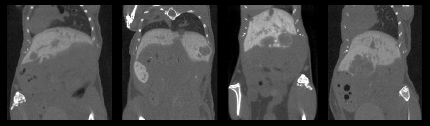
Contrast enhanced CT to detect liver tumors in a HCC mouse model
The X-CUBE is ultimately suited for contrast enhanced anatomical studies in both rat and mouse. In this case, contrast enhanced CT was done to evaluate liver tumors in a hepatocellular carcinoma (HCC) mouse model. For more contrast CT-images we like to refer to the applications page.
Mice were injected with Exitron 12000 at day -1, a nanoparticle-based contrast agent that accumulates in the liver. At day 0, the mice were evaluated with a high resolution CT scan taken on the X-CUBE. The data were reconstructed using the ISRA algorithm, with a voxel size of 100 µm.
Exitron 12000 enhanced CT can also be used to follow up liver tumors longitudinally.
Courtesy of L. Devisscher and S. Van Campenhout et al., Dpt. of Gastroenterology&Hepatology, Hepatology Research Unit, Ghent University