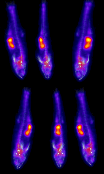
Zebrafish PET-CT imaging
The zebrafish (Danio rerio) is a frequently used animal model to study human disease, since the majority of molecular and cellular mechanisms are conserved between zebrafish and human. In addition, mutations can be introduced easily to mimic human disease.
This study highlights the sub-millimeter resolution and excellent sensitivity of the β-CUBE . 1 MBq (37µCi) of [18F]-NaF was injected in a zebrafish (1 inch/2.5 cm), scanned for 5 min and coregistered with a 3 min high resolution CT acquisition. Even though very short acquisition times combined with a relatively low dose (1MBq/37µCi) were used, bone uptake is clearly visible in the PET image, mainly in the skull and spine. This unprecedented combination of resolution and sensitivity opens up new capabilities to accelerate your translational research workflow by enabling very clear PET imaging of Zebrafish.
This work was done in collaboratIon with the Center for Medical Genetics Ghent and the nuclear medicine and molecular imaging department at UMC Groningen.
