Cardiac gated PET imaging in mouse and rat
Research question
In cardiac research, non-invasive measurement of left ventricular end-systolic (ESV), end-diastolic volume (EDV) and left ventricular ejection fraction (LVEF) is a useful tool to evaluate and monitor cardiac function in health and disease.
Experiment
In order to obtain physiological signals, Molecubes developed dedicated animal beds for mice and rats with fully integrated cardiac, respiratory and temperature monitoring capabilities. The electronics are equipped with a high precision clock to allow synchronisation of the monitoring signals with the CUBES. Three ECG-leads are connected to the front legs and left hind leg to obtain the cardiac signal.
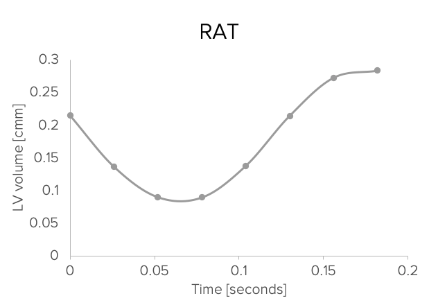
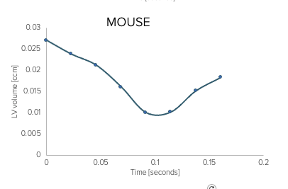
In the examples below, a rat and mouse were injected with 17 and 8.8 MBq, respectively, of [18F]-FDG ± 80 minutes prior to cardiac gated PET scanning (dose at start scan: 10.2 and 5.5 MBq, respectively). For each cardiac cycle, 8 gates were defined and for each gate a PET image was reconstructed using the OSEM algorithm with default settings as provided by the Molecubes software. ESV, EDV and LVEF were calculated using PMOD PCARD v4.0.
Results
Thanks to the high spatial resolution of the PET system, cardiac function can be visualised and quantified in both rats and mice.