Heterocellular 3D scaffolds as biomimetic to recapitulate the tumor microenvironment of peritoneal metastases in vitro and in vivo.
Research question
Peritoneal metastasis is a major cause of death and preclinical models are urgently needed to enhance therapeutic progress. This study reports on a hybrid hydrogel-polylactic acid (PLA) scaffold that mimics the architecture of peritoneal metastases at the qualitative, quantitative and spatial level.
Experiment
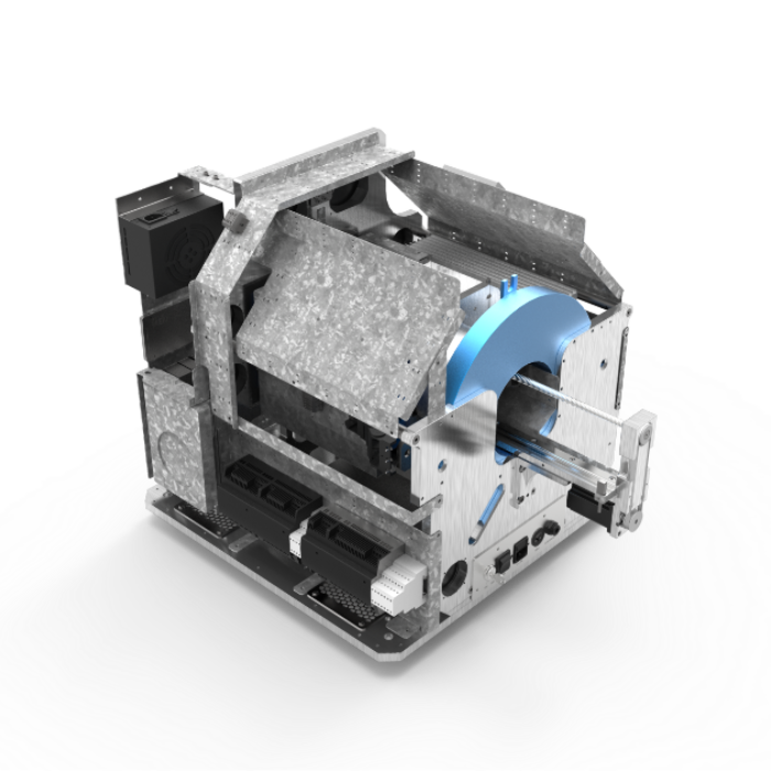
In this paper, the MOLECUBES X-CUBE was used to acquire in vivo contrast enhanced high-resolution CT images of blood vessel infiltration in a non-invasive manner.
Results
Porous PLA scaffolds with controllable pore size, geometry and surface properties are functionalized by type I collagen hydrogel. Co-seeding of cancer-associated fibroblasts (CAF) increases cancer cell adhesion, recovery and exponential growth by in situ heterocellular spheroid formation. Scaffold implantation into the peritoneum allows long-term follow-up (>14 weeks) and results in a time-dependent increase in vascularization, which correlates with cancer cell colonization in vivo. CAF, endothelial cells, macrophages and cancer cells show spatial and quantitative aspects as similarly observed in patient-derived peritoneal metastases. CAF provide long-term secretion of complementary paracrine factors implicated in spheroid formation in vitro as well as in recruitment and organization of host cells in vivo. In conclusion, the multifaceted heterocellular interactions that occur within peritoneal metastases are reproduced in this tissue-engineered implantable scaffold model.
