Imaging CAR T Cell Trafficking with eDHFR as a PET Reporter Gene
In this paper by Sellmyer et al., the MOLECUBES β-CUBE and X-CUBE were used to investigate the ability of [18F]-TMP, a newly developed fluorine-18 probe, to image primary human chimeric antigen receptor (CAR) T cells expressing the PET reporter gene eDHFR.
Research question
Cell-based therapeutics have considerable promise across diverse medical specialties; however, reliable human imaging of the distribution and trafficking of genetically engineered cells remains a challenge.
Experiment
The researchers developed positron emission tomography (PET) probes based on the small-molecule antibiotic trimethoprim (TMP) that can be used to image the expression of the Escherichia coli dihydrofolate reductase enzyme (eDHFR) and tested the ability of [ 18F]-TMP, a fluorine-18 probe, to image primary human chimeric antigen receptor (CAR) T cells expressing the PET reporter gene eDHFR, yellow fluorescent protein (YFP), and Renilla luciferase (rLuc).
Results
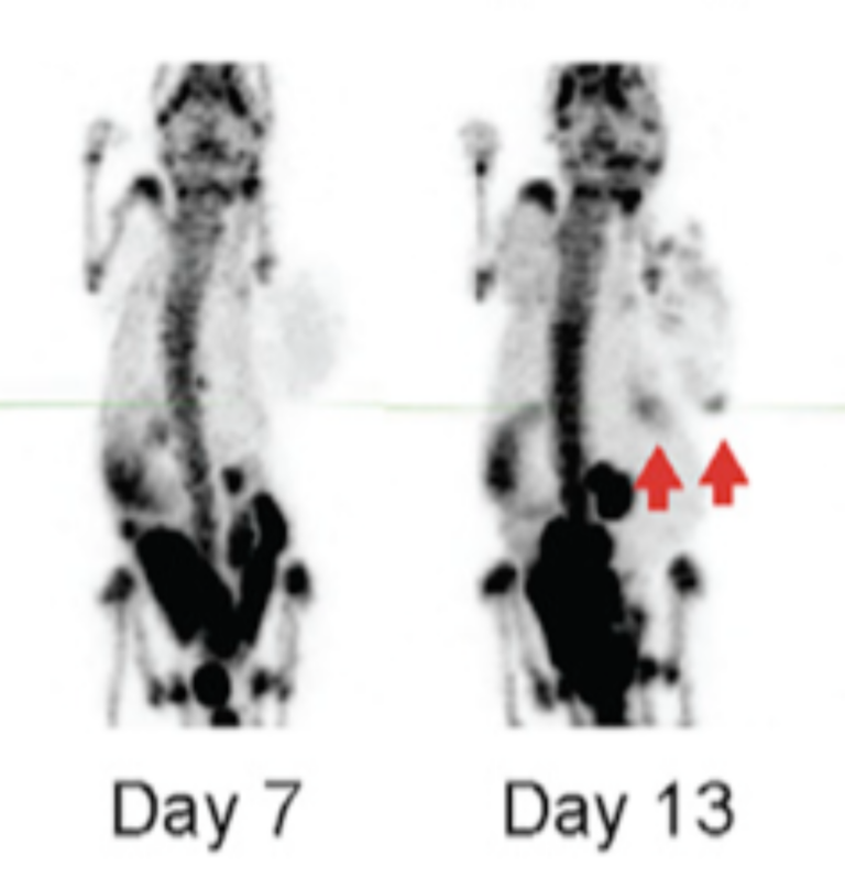
Engineered T cells showed an approximately 50-fold increased bioluminescent imaging signal and 10-fold increased [ 18F]-TMP uptake compared to controls in vitro. eDHFR-expressing anti-GD2 CAR T cells were then injected into mice bearing control GD2 − and GD2 + tumors. PET/CT images acquired on days 7 and 13 demonstrated early residency of CAR T cells in the spleen followed by on-target redistribution to the GD2 + tumors. This was corroborated by autoradiography and anti-human CD8 immunohistochemistry. We found a high sensitivity of detection for identifying tumor-infiltrating CD8 CAR T cells, ∼11,000 cells per mm 3. These data suggest that the [ 18F]-TMP/eDHFR PET pair offers important advantages that could better allow investigators to monitor immune cell trafficking to tumors in patients.