In vivo biodistribution of calcium phosphate nanoparticles after intravascular, intramuscular, intratumoral, and soft tissue administration in mice investigated by small animal PET/CT
In this paper by Kollenda et al., the MOLECUBES β-CUBE and X-CUBE were used to compare the biodistribution of 68Ga-labelled nanoparticles following different application routes: intravenous, intramuscular, intratumoral, and into soft tissue.
Research question
Nanoparticles based on calcium phosphate have become a versatile delivery system for many cargo molecules, including small molecules, DNA, RNA, proteins and antigens for in vitro and in vivo applications. However, the in vivo biodistribution of calcium phosphate nanoparticles still remains unclear as they are difficult to trace inside the body. Calcium phosphate nanoparticles were covalently surface-functionalized with the ligand DOTA and loaded with the radioisotope 68Ga. The aim of this project was to visualize the biodistribution of potentially therapeutic calcium phosphate nanoparticles in vivo by dynamic PET imaging of several application routes.
Experiment
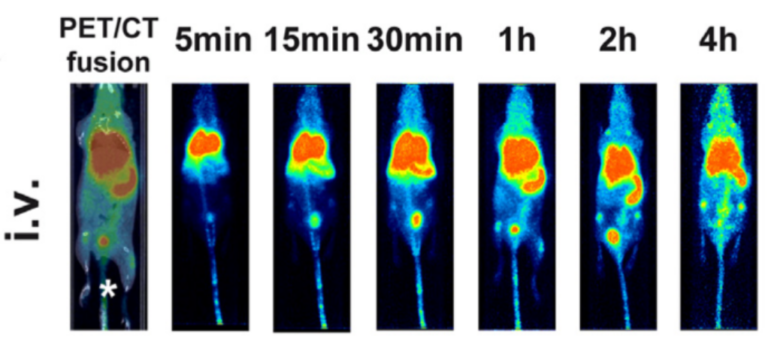
The biodistribution of such 68Ga-labelled nanoparticles was followed in vivo in mice by positron emission tomography in combination with computer tomography (PET-CT). The biodistribution of 68Ga-labelled nanoparticles was compared for different application routes: intravenous, intramuscular, intratumoral, and into soft tissue. The particle distribution was measured in vivo by PET-CT after 5 min, 15 min, 30 min, 1 h, 2 h, and 4 h, and ex vivo after 5 h.
Results
After intravenous injection (tail vein), the nanoparticles rapidly entered the lungs with later redistribution into liver and spleen. The nanoparticles remained mostly at the injection site following intramuscular, intratumoral, or soft tissue application, with less than 10 percent being mobilized into the blood stream.