Increased uptake of the P2X7 receptor radiotracer 18 F-JNJ-64413739 in the brain and peripheral organs according to the severity of status epilepticus in male mice
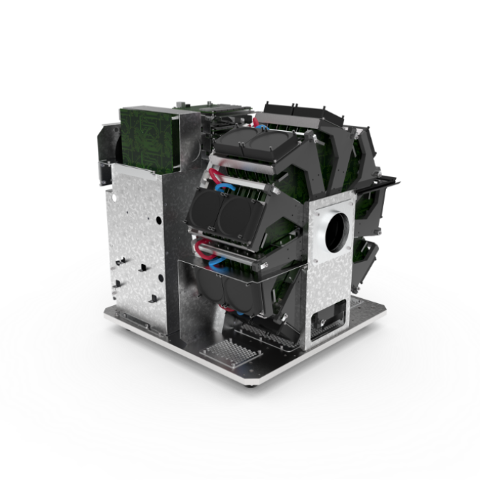
In this study by Morgan et al., the β‐CUBE and X‐CUBE were used as a non-invasive tool for the identification and evaluation of disease‐specific biomarkers. The cubes allowed for the longitudinal evaluation of a potential non-invasive biomarker of epilepsy.
Research question
Adenosine triphosphate (ATP) acts as a co-transmitter to regulate brain network excitability via the activation of purinergic P2 receptors, including the ligand-gated cationic P2X receptors. Among these, the P2X7 receptor (P2X7R) has attracted attention as a potential drug target and biomarker for epilepsy. P2X7R is involved in numerous pathways relevant to epileptogenesis, the process of turning a healthy brain into a brain experiencing epileptic seizures, including synaptic plasticity, neurogenesis, and the regulation of neurotransmitter release. Genetic or pharmacologic targeting of P2X7R can reduce seizure frequency and severity in preclinical models. Although P2X7R radiotracer uptake has successfully been detected in the brain of rodents and humans, whether altered P2X7R expression following status epilepticus (SE) can be detected via PET and whether this signal reflects underlying pathology relevant to epilepsy remains unknown. In this study, 18F-JNJ-64413739, a positron emission tomography (PET) P2X7R antagonist, was tested as a potential non-invasive biomarker of seizure damage and epileptogenesis.
Experiment
Status epilepticus was induced in mice via an intra-amygdala microinjection of kainic acid. Static PET studies (30min duration, initiated 30min after tracer administration) were conducted 48h after status epilepticus via an intravenous injection of 18F-JNJ-64413739. PET images were co-registered with a brain magnetic resonance imaging atlas, tracer uptake was determined in the different brain regions and peripheral organs, and values were correlated to seizure severity during status epilepticus. 18F-JNJ-64413739 was also applied to ex vivo human brain slices obtained following surgical resection for intractable temporal lobe epilepsy.
Results
Using a mouse model of SE leading to drug‐resistant focal epilepsy, this study shows acute elevation in signal from the P2X7R‐specific radiotracer 18F‐JNJ‐64413739 according to the severity of SE in the brain and peripheral organs. P2X7R radiotracer uptake correlated strongly with seizure severity during status epilepticus in brain structures including the cerebellum and ipsi- and contralateral cortex, hippocampus, striatum, and thalamus. In addition, a correlation between radiotracer uptake and seizure severity was also evident in peripheral organs such as the heart and the liver. P2X7R radiotracer uptake levels in the brain and peripheral organs correlate with the severity of status epilepticus. Finally, P2X7R radiotracer uptake was found elevated in brain sections from patients with temporal lobe epilepsy when compared to control.
This study provided the first evidence of the diagnostic potential of P2X7R‐based PET imaging for seizure‐induced pathology and epilepsy. Importantly, with the demonstrated role of P2X7R during seizures and epilepsy, P2X7R PET imaging may represent a new diagnostic tool for patients according to seizure-induced neuropathology and temporal lobe epilepsy. The findings of this study suggest that PET imaging of P2X7R may help to identify patients with increased P2X7R expression and, hence, in need of P2X7R‐based treatments.

