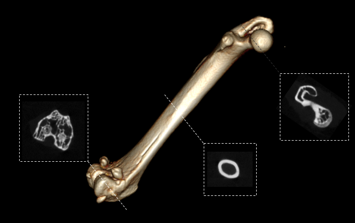
Mouse femur imaging
Preclinical models for musculoskeletal disorders are important for understanding the pathogenesis of bone and joint disorders in humans and to develop effective therapies. In vivo bone imaging studies on the X-CUBE allow the researcher to evaluate specific bones longitudinally in a non-invasive manner.
The image shown below displays a mouse femur. Postprocessing software can be used to calculate BV/TV, cortical thickness and surface area, among others.