Testimonial Prof. Dr. Christian Vanhove (BE)
Prof. Christian Vanhove is responsible for the day-to-day functioning of the preclinical INFINITY lab (Ghent, Belgium) and the coordination of the partner research, either from within Ghent University, from private pharma or biotech companies. He has a MOLECUBES PET/SPECT/CT benchtop combination operational in the lab since mid 2016.
Why choose MOLECUBES?
The lab will move to a new building in a few years from now. Luckily, the benchtop modular system allows me to move all 3 modalities at no extra cost or time loss for recalibration. The new lab will have less space, so I’m very happy that these benchtop systems exist today without compromise on image quality.
How would your rate your experience with the MOLECUBES systems?
The PET, SPECT and CT hardware and software proved to be very robust and reliable over the past year. To date, we have already imaged over 100 mice and rats effortlessly. Whenever we have a new request from a partnering lab, we can reach out to Molecubes for questions related to the specific projects.
What specific features do you like most?
All 3 modalities provide 1 convenient DICOM file to work with, there is no manual interaction needed for co-registration and still all CUBES can be used in stand-alone mode when needed. The whole process is automated and produces publication ready images. To me, that’s true flexibility!
EXAMPLE PROJECTS
These studies were performed by PhD students and required minimal training efforts from the team.
PET CT to evaluate bone metastasis
Protocol
full body rat (200g)- 3 bed positions; 10 min/bed position – 10.67 MBq [18F]FDG
– 30 min uptake
OSEM reconstruction: 50 iterations, 400µm voxel size HR spiral CT, 7 min acquisition ISRA reconstruction – 200 um voxel size
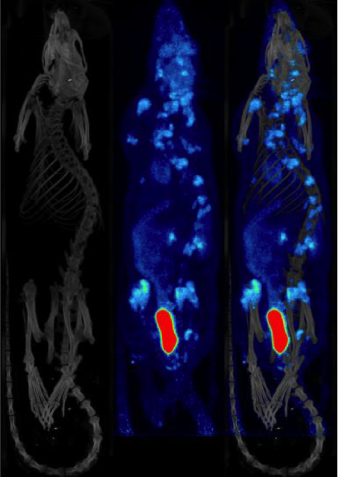
Courtesy of Valerie Demeulenaere et al., Department of Radiology, Ghent University Hospital
PET MR – Rat with glioblastoma
Protocol
Rat with glioblastoma – 1 bed position; 10 min acquisition time
– 8.89 MBq [18F]FDG – 1h30 uptake – OSEM reconstruction: 50 iterations, 400µm voxel size – Gadolineum enhanced T1 weighted image, 7T system (Pharmascan, Bruker)
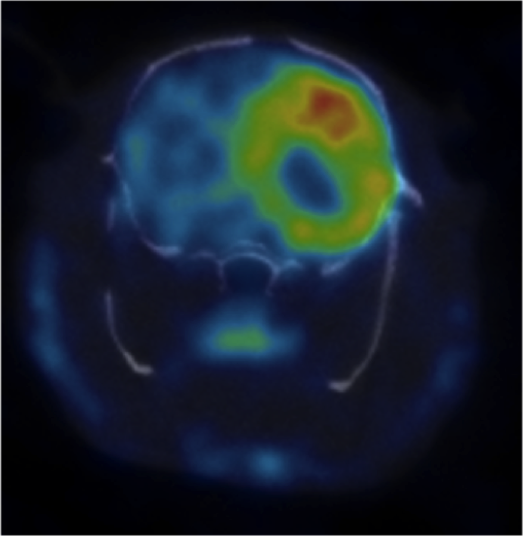
Courtesy of Julie Bolcaen et al., Department of Nuclear Medicine, Ghent University Hospital